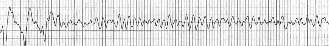
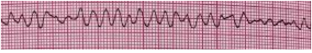
Coarse V Fib Ecg
Ventricular Fibrillation Vf Litfl Ecg Library Diagnosis
Ventricular Fibrillation Vf Litfl Ecg Library Diagnosis
Ventricular Fibrillation Vf Litfl Ecg Library Diagnosis
Ventricular Fibrillation Vf Litfl Ecg Library Diagnosis

Ventricular Fibrillation Cmwtfm Presents Fsm

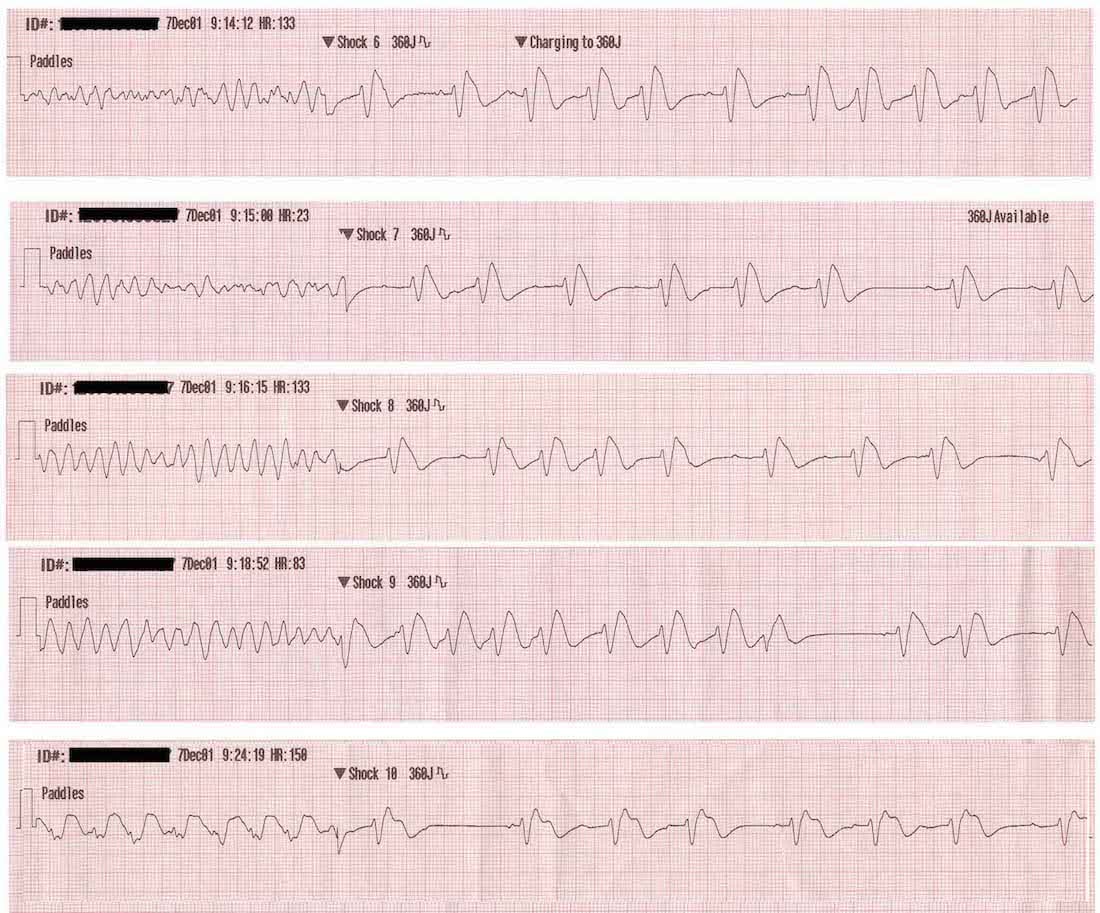
Acls Ventricular Fibrillation And Pulseless Ventricular Tachycardia Guide

These features include observing p wave forms measurement of ekg intervals and segments assessment of rhythm calculating heart rate and the evaluation of other relevant wave segments.
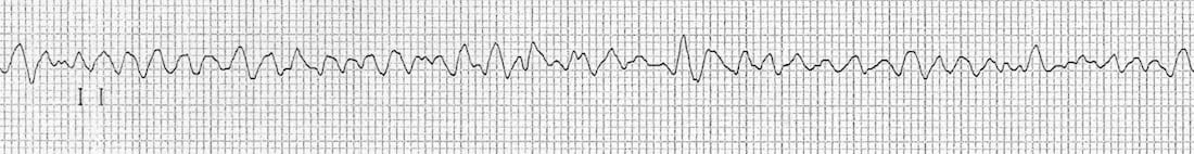
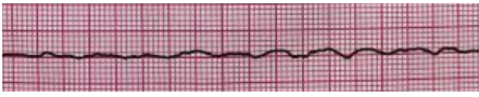
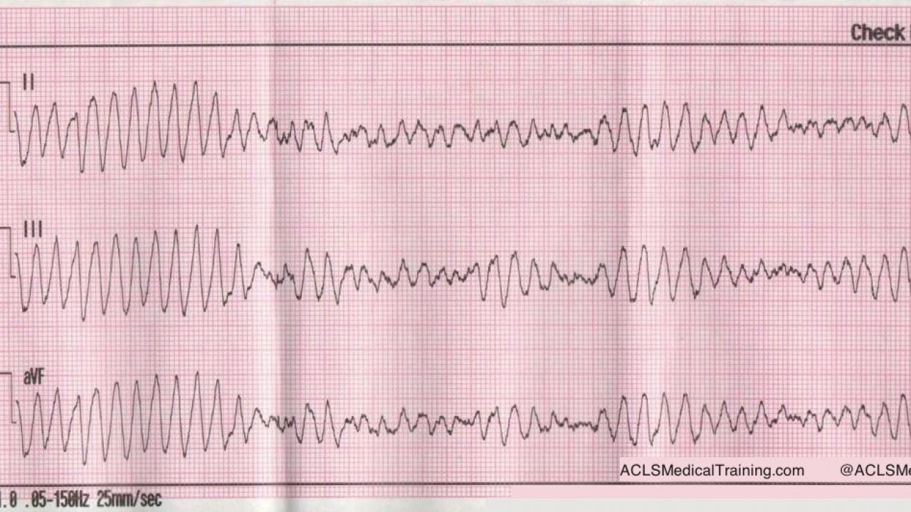
Coarse v fib ecg. In addition the 6 lead ecg recorded during early vf had episodes of coarse vf and fine vf occurring simultaneously. Mother rotor mechanism in which a stable re entry circuit is formed the mother rotor. A good starting point for learning about v fib and other types of ekg interpretation is our ekg basics training course. Fine ventricular fibrillation is even more dangerous than coarse ventricular fibrillation because there is even less contractility of the myocardium which results in a smaller amplitude.
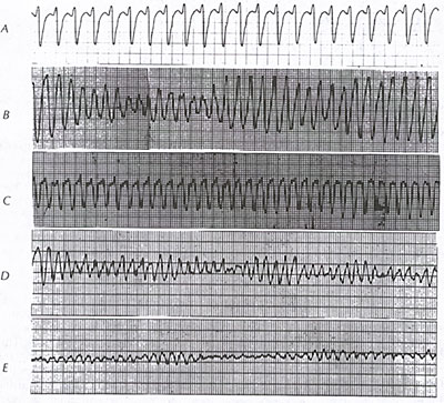
Usually when a person experiences ventricular fibrillation it first starts off as coarse ventricular fibrillation. Coarse af indicates atrial enlargement. 03 10 ventricular fibrillation v fib 03 11 1st degree av heart block 03 12 2nd degree av heart block type 1 mobitz i wenckebach. Fine v fib is sometimes mistaken for asystole.
Coarse ventricular fibrillation is more likely to convert after defibrillation than fine v fib. In this atrial fibrillation ecg review the ecg criteria to diagnose atrial fibrillation afib including atrial fibrillation with rvr coarse atrial fibrillation and other af scenarios are. Once those characteristics are identified the next thing to look for is the absence of p waves and an undulating baseline that can appear almost flat fine or very rough coarse. Such types of atrial fibrillation is likely to be persistent unless the cause of atrial dilatation is reversible with an intervention like balloon mitral valvotomy.
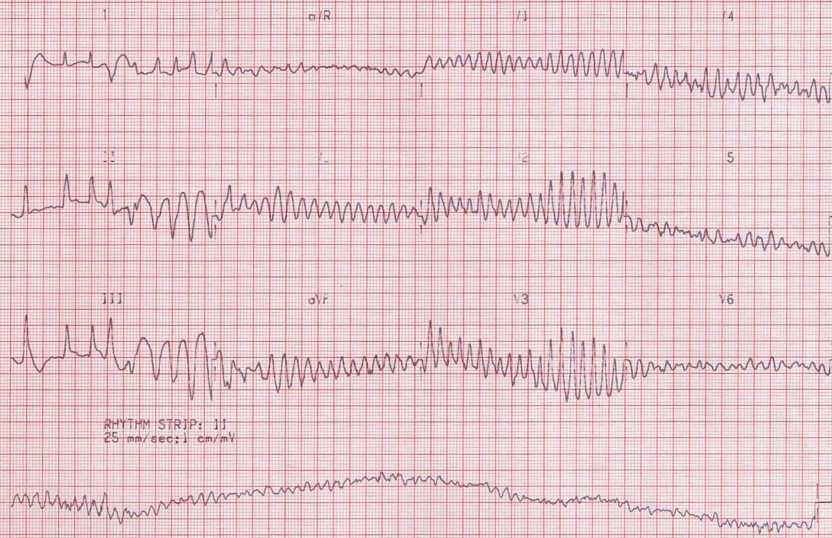
We conclude that coarse vf does not reflect greater synchronization of activity than fine vf early in fibrillation five minutes. The fibrillation is maintained by re entry circuits formed by some of the wavelets. Ventricular fibrillation may be fine or coarse. The mother rotor then gives rise to propagating unstable daughter wavefronts which results in the chaotic electrical activity seen on the ecg.
In dogs and cats atrial fibrillation is characterized on the ecg by a supraventricular normal appearing qrs complexes and an irregular ventricular rhythm that is most commonly fast. As the treatments for asystole and ventricular fibrillation are different it is important to differentiate between the two.
Ventricular Fibrillation

Ventricular Fibrillation Wikipedia
Section 9 Ventricular Rhythm

A 10 Coarse Ventricular Fibrillation Mixed With Ventricular Flutter Download Scientific Diagram
Ventricular Fibrillation Vf Litfl Ecg Library Diagnosis
Difference Between Coarse And Fine Ventricular Fibrillation

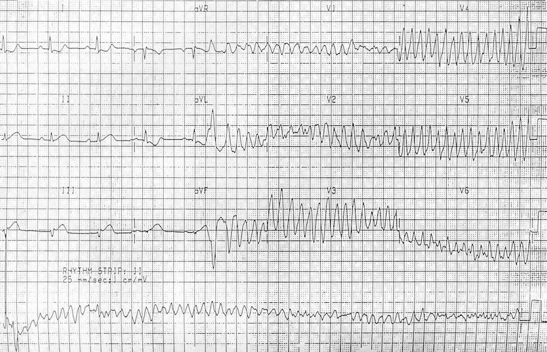
Dr Smith S Ecg Blog Ventricular Fibrillation On A 12 Lead Ecg

Matters Of The Heart Cardiac Arrest Patmac Rn
Difference Between Coarse And Fine Ventricular Fibrillation
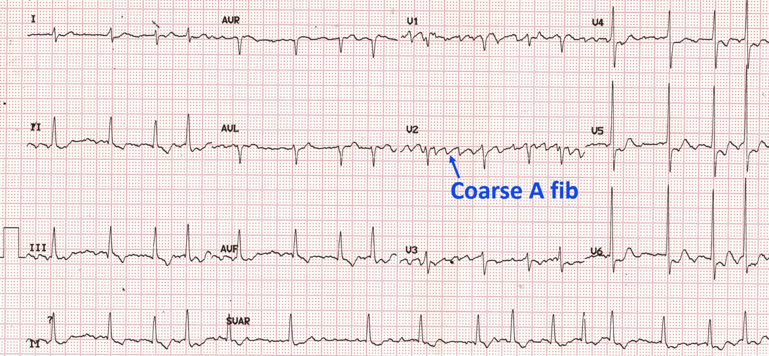
Coarse Atrial Fibrillation On Ecg All About Cardiovascular System And Disorders

Pin On School Stuff
What Is Fine V Fib And What Makes It Different From V Fib Quora
Cpr First Or Defibrillation First Acls Medical Training
